2. 宁夏医科大学第三临床医学院, 银川 750004
2. The Third Clinical Medical College of Ningxia Medical University, Yinchuan 750004, Ningxia Hui Autonomous Region, China
胃息肉是常见的起源于胃黏膜上皮或黏膜下的局限性隆起样病变。绝大部分单纯胃息肉患者无特异性临床症状,合并其他消化道疾病时表现为腹痛、腹胀、反流、消化不良、消化道出血,发生在贲门或幽门部时可导致消化道梗阻,常在上消化道内镜检查中被偶然发现[1]。胃息肉按照病理类型可分为腺瘤性息肉、增生性息肉、炎性息肉、错构瘤性息肉及其他类型。本研究就宁夏人民医院2012-2019年检出的胃息肉进行相关报道。
1 资料与方法 1.1 一般资料选取2012年10月至2019年10月宁夏人民医院收治的行内镜检查患者90 643例,胃镜下诊断为胃息肉者共3 807例,男性1 434例,女性2 373例,检出率为4.2%,年龄26~84岁,平均年龄为(54.03±12.18)岁,其中行病理学检查1 774例,除去35例确诊为肿瘤者,病理结果明确诊断为胃息肉者共1 170例,平均年龄为(53.02±11.53)岁,以女性较为常见(男女比例为1.7∶1)。
纳入标准:胃内准备充分,不影响胃黏膜的观察;胃镜完成对胃内各部位观察;经过活检病理或电凝切除后病理检查证实为胃息肉。排除标准:有胃手术史;胃息肉病;胃内准备不良影响黏膜观察;胃镜未能完成患者。所有患者均知情且签署知情同意书。
1.2 胃镜检查方法行胃镜检查前告知患者可能出现的风险及并发症,征得患者同意并签署知情同意书,由经验丰富的内镜医生对患者的食管、胃及十二指肠反复查看,并记录胃息肉的数量、部位、大小等信息,对部分患者检测组织病理学检查及幽门螺杆菌(Hp)感染情况。
1.3 统计学处理采用SPSS 23.0软件行统计学分析,符合正态分布的计量资料以x±s表示,采用方差分析,计数资料以n(%)表示,采用χ2检验比较组间差异。检验水准(α)为0.05。
2 结果 2.1 不同病理类型胃息肉患者的一般资料分析结果(表 1)显示:病理结果中最常见的类型依次为胃底腺息肉410例(32.3%)、增生性息肉351例(29.3%)、炎性息肉287例(22.6%)、腺瘤性息肉120例(9.5%)、错构瘤性息肉2例(0.2%)。其中各个病理类型的息肉在不同性别间差异有统计学意义(P < 0.05),除腺瘤性息肉患者中男性较女性多,所有息肉类型均是女性数量较多,尤其是炎性息肉(2.90∶1)。不同病理类型的息肉均在40~60岁多发,但在年龄方面差异无统计学意义(P=0.09)。胃底腺息肉多发较单发稍常见,其余均以单发为主(P < 0.05),腺瘤性息肉的大小多分布在5 mm以上,其余病理类型息肉以5 mm以下多见(P < 0.05)。所有的病理类型的患者均以胃体常见(P < 0.05)。
| n(%) | |||||||||||||||||||||||||||||
| 指标 | 腺瘤性息肉组(n=120) | 增生性息肉组(n=351) | 炎性息肉组(n=287) | 胃底腺息肉组(n=410) | χ2值 | P值 | |||||||||||||||||||||||
| 性别 | 36.701 | < 0.000 1 | |||||||||||||||||||||||||||
| 男 | 63(52.5) | 139(39.6) | 91(31.7) | 105(25.6) | |||||||||||||||||||||||||
| 女 | 57(47.5) | 212(60.4) | 196(68.3) | 305(74.4) | |||||||||||||||||||||||||
| 年龄/岁 | 22.745 | 0.090 | |||||||||||||||||||||||||||
| ≤30 | 2(1.7) | 11(3.2) | 12(4.2) | 13(3.2) | |||||||||||||||||||||||||
| 31~40 | 11(9.2) | 36(10.3) | 25(8.7) | 48(11.7) | |||||||||||||||||||||||||
| 41~50 | 25(20.8) | 89(25.4) | 79(27.5) | 125(30.5) | |||||||||||||||||||||||||
| 51~60 | 34(28.3) | 118(33.6) | 100(34.8) | 114(27.8) | |||||||||||||||||||||||||
| 61~70 | 42(35.0) | 76(21.7) | 55(19.2) | 82(20.0) | |||||||||||||||||||||||||
| >70 | 6(5.0) | 21(6.0) | 16(5.6) | 28(6.8) | |||||||||||||||||||||||||
| 数量 | 12.745 | 0.005 | |||||||||||||||||||||||||||
| 单发 | 64(53.3) | 196(55.8) | 178(62.0) | 199(48.5) | |||||||||||||||||||||||||
| 多发 | 56(46.7) | 155(44.2) | 109(38.0) | 211(51.5) | |||||||||||||||||||||||||
| 大小/mm | 117.035 | < 0.000 1 | |||||||||||||||||||||||||||
| ≤5 | 50(41.7) | 242(68.9) | 215(74.9) | 312(76.1) | |||||||||||||||||||||||||
| 5~10 | 36(30.0) | 95(27.1) | 58(20.2) | 81(19.8) | |||||||||||||||||||||||||
| >10 | 34(28.3) | 14(4.0) | 14(4.9) | 17(4.1) | |||||||||||||||||||||||||
| 部位 | 82.146 | <0.000 1 | |||||||||||||||||||||||||||
| 贲门/胃底 | 27(22.5) | 73(20.8) | 62(21.6) | 107(26.1) | |||||||||||||||||||||||||
| 胃体 | 51(42.5) | 153(43.7) | 126(43.9) | 206(50.2) | |||||||||||||||||||||||||
| 胃窦/幽门/胃角 | 21(17.5) | 55(15.7) | 61(21.3) | 4(1.0) | |||||||||||||||||||||||||
| 多部位 | 21(17.5) | 70(19.9) | 38(13.2) | 93(22.7) | |||||||||||||||||||||||||
| n(%) | |||||||||||||||||||||||||||||
| 指标 | 腺瘤性息肉组(n=120) | 增生性息肉组(n=351) | 炎性息肉组(n=287) | 胃底腺息肉组(n=410) | χ2值 | P值 | |||||||||||||||||||||||
| Hp感染 | 11(9.2) | 98(27.9) | 92(32.0) | 33(8.3) | 85.125 | < 0.05 | |||||||||||||||||||||||
| 肠化 | 11(9.2) | 28(8.0) | 30(10.5) | 25(6.1) | 4.556 | 0.207 | |||||||||||||||||||||||
| 活动性炎症 | 5(4.2) | 23(6.6) | 20(7.0) | 20(3.9) | 2.296 | 0.513 | |||||||||||||||||||||||
| 非典型增生 | 29(24.2) | 9(2.6) | 11(3.8) | 8(2.0) | 108.469 | < 0.05 | |||||||||||||||||||||||
在不同病理类型的的胃息肉中,经过对幽门螺杆菌感染的比较,增生性息肉及炎性息肉患者Hp感染率较高,分别为27.9%和32.0%,胃底腺息肉及腺瘤性息肉的感染率较低,为8.3%和9.2%,且差异具有统计学意义(P < 0.05)。非典型增生的发生率在腺瘤性息肉明显高于其他类型息肉(P < 0.05),不同类型的息肉在肠化及活动性炎症方面,差异无统计学意义。
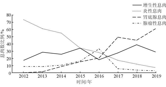
2.3 胃息肉病理类型比例的变化趋势结果(图 1)显示:近7年内,增生性息肉占30%左右,腺瘤性息肉在2016年达高峰,为33.3%,其余在10%左右,胃底腺息肉比例升高至50.2%,炎性息肉比例从64.5%下降至10.5%。

|
| 图 1 2012-2019年不同病理类型胃息肉占比的变化趋势 |
胃息肉是一种突出于胃黏膜表面的良性隆起性病变,年龄、遗传、环境因素、Hp感染、长期使用质子泵抑制剂、萎缩性胃炎等都影响胃息肉的形成。对于临床上有症状的息肉,常采用胃镜下干预及治疗,而大部分无症状息肉,是否进行干预及治疗,还需根据内镜检查及病理学检查的相关特点进行进一步评估。
本研究分析2012-2019年宁夏地区1 170例胃息肉患者病例资料发现,在我院就诊患者的常见胃息肉病理类型发生了改变,胃底腺息肉已代替增生性息肉成为目前最常见的胃息肉类型。国外研究[1]表明,近年来增生性息肉比例较前明显下降,胃底腺性息肉已成为目前最为常见的胃息肉病理类型。国内的一项对10 137例胃息肉患者的研究[2]也得出类似结果。Hp的感染和增生性息肉关系密切,考虑近年来随着人们生活习惯的改善和治疗意识的提高,Hp感染率有所降低,增生性息肉的比例下降可能与此有关。胃底腺息肉生成与质子泵抑制剂(PPI)类药物的使用密切相关,主要是通过抑制壁细胞分泌H+而达到抑酸作用,壁细胞排H+不畅而肿胀凸起,导致胃底腺堵塞,从而引起胃黏膜隆起,部分患者停用PPI类药物后,胃底腺息肉可自行消退[3]。但因本研究是回顾性分析,对患者PPI类药物的服用情况不明确,与之是否有关,还需进一步前瞻性研究证实。
从一般的人口学特征来看,胃息肉除在腺瘤性息肉中男性较多见外,其余类型均多发于女性,这与国内外的相关报道[2, 4-5]一致。胃息肉患者年龄大多分布在40~60岁,腺瘤性息肉在60岁以上的患者比例约为40%,明显高于其他病理类型,年龄是腺瘤性息肉的危险因素之一[6],且本研究中,腺瘤性息肉的非典型增生发生率较其他类型息肉明显高,不同病理类型的胃息肉具有明显不同的恶变潜能,胃底腺息肉几乎无恶变风险,增生息肉极少发生恶变,腺瘤性息肉的癌变率明显高于其他类型[7]。从胃息肉的大小方面来说,除腺瘤性息肉以外,均以5 mm以下多发,相关研究[8]表明,大于10 mm应当尽早切除。本研究中10 mm以上的以癌变率较高的腺瘤性息肉多见,因此,60岁以上无症状男性患者胃镜发现有息肉,且息肉大小超过10 mm时,应当更加倾向考虑给与胃镜下治疗及干预,以免日后发生癌变。
Hp感染是慢性胃炎的常见原因之一,胃黏膜在长期慢性炎症的刺激下,胃上皮细胞增生,导致息肉形成。本研究中胃息肉的总体Hp的感染率为20.0%,与国内外相关报道[9-11]大致类似,其中以增生性息肉及炎性息肉的Hp感染率最高。相关研究[11-12]表明,Hp感染和增生性息肉及炎性息肉关系密切。Hp感染可导致胃黏膜的肠化、异型性增生,有进一步增大和癌变倾向[13-14]。且相关研究[15]表明Hp感染根治后可减少息肉发生,指南[16]推荐增生息肉合并Hp感染时应进行根除治疗,考虑到Hp感染与胃息肉的关系,对增生性息肉及炎性息肉合并Hp感染患者应当积极根除Hp,定期复查胃镜。
相关研究[17]表明,除腺瘤性息肉外,其余胃息肉大多都集中在胃体、胃底。本研究中息肉均以胃体部为高发,误差可能由于腺瘤性息肉样本量较小,因而差异无统计学意义。本研究发现胃底腺息肉常见多发,其余均多为单发,这与大部分研究[2, 4]相吻合。
综上所述,对于胃息肉的研究仍有不少争议,不同地区和人群,不同类型的胃息肉有各自的特点。鉴于近年来胃息肉谱发生变化,恶变可能性较小的胃底腺息肉的比例增高,因此,对胃息肉进行切除时,应当对其一般的特征、病理学特征及是否合并Hp感染进行比较分析后作出相应决定。
利益冲突:所有作者声明不存在利益冲突。
| [1] |
JAIN R, CHETTY R. Gastric hyperplastic polyps: a review[J]. Dig Dis Sci, 2009, 54(9): 1839-1846.
[DOI]
|
| [2] |
何金龙, 陈磊, 代剑华, 等. 10137例胃息肉的临床及病理特征分析[J]. 第三军医大学学报, 2018, 40(3): 248-254. HE J L, CHEN L, DAI J H, et al. Clinical and pathological characteristics of gastric polyps: a report of 10137 patients[J]. Journal of third military medical university, 2018, 40(3): 248-254. [CNKI] |
| [3] |
TRAN-DUY A, SPAETGENS B, HOES A W, et al. Use of proton pump inhibitors and risks of fundic gland polyps and gastric cancer: systematic review and meta-analysis[J]. Clin Gastroenterol Hepatol, 2016, 14(12): 1706-1719.e5.
[DOI]
|
| [4] |
ROMAN M A, AGNIESZKA M, KATARZYNA G U. Pathophysiological and clinical aspects of gastric hyperplastic polyps[J]. World J Gastroenterol, 2016, 22(40): 8883-8891.
[DOI]
|
| [5] |
FAN N N, YANG J, SUN G, et al. Changes in the spectrum of gastric polyps in the Chinese population[J]. World J Gastroenterol, 2015, 21(33): 9758-9764.
[DOI]
|
| [6] |
蔡欧, 俞媛洁, 袁静萍, 等. 胃小息肉中腺瘤性息肉危险因素的临床研究[J]. 胃肠病学, 2018, 23(7): 426-428. CAI O, YU Y J, YUAN J P, et al. Risk factors of adenomatous polyp in small gastric polyps[J]. Chinese Journal of Gastroenterology, 2018, 23(7): 426-428. [DOI] |
| [7] |
ASGE Standards of Practice Committee, EVANS J A, CHANDRASEKHARA V, et al. The role of endoscopy in the management of premalignant and malignant conditions of the stomach[J]. Gastrointest Endosc, 2015, 82(1): 1-8.
[DOI]
|
| [8] |
SHAIB Y H, RUGGE M, GRAHAM D Y, et al. Management of gastric polyps: an endoscopy-based approach[J]. Clin Gastroenterol Hepatol, 2013, 11(11): 1374-1384.
[DOI]
|
| [9] |
JI F, WANG Z W, NING J W, et al. Effect of drug treatment on hyperplastic gastric polyps infected with Helicobacter pylori: a randomized, controlled trial[J]. World J Gastroenterol, 2006, 12(11): 1770-1773.
[DOI]
|
| [10] |
代飞, 郑义, 徐丽红, 等. 胃息肉临床特点及其与幽门螺杆菌感染的相关性研究[J]. 胃肠病学和肝病学杂志, 2017, 26(6): 686-688. DAI F, ZHENG Y, XU L H, et al. Clinical characteristics of gastric polyps and the relationship with Helicobacter pylori infection[J]. Chinese Journal of Gastroenterology and Hepatology, 2017, 26(6): 686-688. [CNKI] |
| [11] |
ELHANAFI S, SAADI M, LOU W, et al. Gastric polyps: association with Helicobacter pylori status and the pathology of the surrounding mucosa, a cross sectional study[J]. World J Gastrointest Endosc, 2015, 7(10): 995-1002.
[DOI]
|
| [12] |
徐玉珍, 费素娟. 三种常见类型胃息肉的分布及其与幽门螺杆菌感染的关系[J]. 胃肠病学, 2018, 23(11): 47-49. XU Y Z, FEI S J. Distribution of three common types of gastric polyps and their correlation with Helicobacter pylori infection[J]. Chinese Journal of Gastroenterology, 2018, 23(11): 47-49. [CNKI] |
| [13] |
MARKOWSKI A R, MARKOWSKA A, GUZINSKA-USTYMOWICZ K. Pathophysiological and clinical aspects of gastric hyperplastic polyps[J]. World J Gastroenterol, 2016, 22(40): 8883-8891.
[DOI]
|
| [14] |
NAKAMURA S, MATSUMOTO T, KOBORI Y, et al. Impact of Helicobacter pylori infection and mucosal atrophy on gastric lesions in patients with familial adenomatous polyposis[J]. Gut, 2002, 51(4): 485-489.
[DOI]
|
| [15] |
SHIMIZU T, MARUSAWA H, WATANABE N, et al. Molecular pathogenesis of Helicobacter pylori-related gastric cancer[J]. Gastroenterol Clin North Am, 2015, 44(3): 625-638.
[DOI]
|
| [16] |
中华医学会消化病学分会幽门螺杆菌和消化性溃疡学组, 全国幽门螺杆菌研究协作组, 刘文忠, 等. 第五次全国幽门螺杆菌感染处理共识报告[J]. 中华消化杂志, 2017, 37(6): 364-378. Helicobacter Pylori and Peptic Ulcer Group of Gastroenterology Branch of Chinese Medical Association, National Helicobacter Pylori Research Cooperation Group, LIU W Z, et al. The fifth national consensus report on the treatment of Helicobacter pylori infection[J]. Chin J Dig, 2017, 37(6): 364-378. [CNKI] |
| [17] |
林泳, 聂玉强, 王红, 等. 近15年2643例胃息肉临床病理学特征和变化趋势分析[J]. 中华消化杂志, 2014, 34(4): 247-250. LIN Y, NIE Y Q, WANG H, et al. Analysis of clinical pathological characteristics and pattern changes of 2643 gastric polyps in the past 15 years[J]. Chin J Dig, 2014, 34(4): 247-250. |
 2021, Vol. 28


