2. 上海联影医疗科技股份有限公司, 上海 200032
2. Shanghai Lianying Medical Technology Co., Ltd., Shanghai 200032, China
冠脉CT造影(coronary CT angiography,CCTA)作为一种无创检查方法能有效排查冠脉疾病[1]。传统影响CCTA的两大关键因素是呼吸和心率。但是心血管病患者年龄偏大,常伴有听力障碍、语言交流困难,或合并呼吸或神经系统疾病等,CT检查过程中实现呼吸控制较为困难,严重影响检查效果。320排探测器CT的优点是具有达16 cm的宽探测器准直,使覆盖整个心脏的扫描在1次轴向旋转中完成且时间短于1 s[2],降低了呼吸运动对CCTA的影响,提高了检查的时间分辨率,是目前CCTA研究的热点。
前期对依从性较差幼儿的研究[3]发现,自由呼吸CCTA与标准屏气CCTA的图像质量无明显差别,双源CT比单源CT更能提高自由呼吸幼儿的冠脉可见度[4]。但目前仍缺乏成人相关的研究结果。上海联影医疗科技股份有限公司研发的uCT960+是目前转速最快的320排CT仪器,其球管旋转时间为0.25 s,结合超高系统转速与时空探测器,能够实现时间分辨率、空间分辨率与对比度分辨率的多重提升。ePhase智能寻心算法能基于各心动周期智能重建和选择最佳时相,提升病情复杂患者的检查成功率。因此,本研究探讨自由呼吸状态下用宽体探测器CT经单心动周期行320排CCTA的可行性,为其临床应用提供参考。
1 资料与方法 1.1 一般资料随机纳入2020年6月至8月临床疑诊或确诊冠脉病变,于我院放射诊断科行CCTA的成年患者100例,最终入组94例,其中男性56例、女性38例,年龄31~85岁,平均年龄(59±12)岁。排除标准:碘对比剂过敏,肾功能不全,甲状腺功能亢进,心律不齐,心率>75次/min[5]。本研究获得医院伦理委员会批准(B2020-061R);患者知情同意。
1.2 CCTA扫描采用320排CT机(uCT960+,联影),探测器16 cm,球管旋转机时间为0.25 s/圈。排除禁忌证后,所选患者CCTA进行检查前1~1.5 h口服美托洛尔25~50 mg,并于检查前舌下含服硝酸甘油0.5 mg。扫描范围为自隆突下至心底心脏区域。扫描前根据患者心电信息进行采集时相的设置,均采用前瞻性心电门控扫描。心率≤65次/min时,采集70%~80% R-R间期;心率为65~75次/min时,采集30%~50% R-R间期。根据扫描方案,将研究对象分为自由呼吸组(n=47)和屏气组(n=47)。两组管电压100~120 kV,管电流120~140 mA,其余扫描参数也相同。
1.3 对比剂注射方案患者均采用双筒高压注射器经右侧肘静脉注射非离子型对比剂碘帕醇注射液(含碘370 g/L),流速4~5 mL/s,注射总量0.7 mL/kg,对比剂注射完后再以同样的流速注入0.9%氯化钠液25 mL。采用Bolus-Tracking自动触发扫描技术,将触发感兴趣区(region of interest,ROI)定于心脏四腔心层面的降主动脉,触发阈值设定为120 HU,当超过触发阈值时机器延迟6 s自动启动CCTA扫描程序。
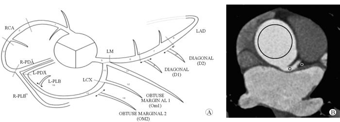
1.4 CT图像后处理将层厚0.5 mm、层间距0.5 mm,使用KARL 3D重建算法、C-SOFT-AA滤波函数,DFOV为220 mm的图像传至2名放射科医师,由其根据美国心脏协会(American Heart Association,AHA)冠脉18节段模型,在主动脉根部、右冠状动脉(RCA)近段及中段、左冠状动脉(LCA)主干、前降支(LAD)近段、回旋支(LCX)近段的ROI中测量CT值和SD。利用信噪比(signal to noise ratio,SNR)和对比噪声比(contrast to noise ratio,CNR)对目标图像质量进行评估[6]。根据心血管计算机体层摄影学会冠脉模型,对1、2、5、6、11节段(左主干、LAD近段、LCX近段、RCA近段及中段)进行分析(图 1A)。ROI位于1、2、5、6、11节段(图 1B)。

|
| 图 1 冠脉测量部位示意图(A)及ROI定位(B) |
采用5分法对冠脉图像质量进行评分[7]。1分:血管模糊不清,伪影严重,无法诊断;2分:血管轮廓模糊,伪影严重,影响诊断;3分:血管轮廓模糊,有明显伪影,不影响诊断;4分:血管轮廓较清晰,伪影较小,不影响诊断;5分:血管轮廓清晰,无伪影,完全能够用于诊断。尽量避开上腔静脉、主动脉壁及肌间隙,选择尽可能大的ROI计算1、2、5、6、11节段的SNR和CNR[8]。SNR=主动脉/背景噪声,CNR=(升主动脉CT值-竖脊肌CT值)/背景噪声,计算冠脉病变的诊断准确性。记录CT扫描系统自动生成的容积CT剂量指数(CTD)、剂量长度乘积(DLP),计算有效辐射剂量(effective dose, ED)。ED=k×DLP,k=0.014 mSv/(mGy×cm)计算[9-11]。
1.6 统计学处理采用SPSS 14.0统计学软件处理数据。采用K-S检验正态分布,符合正态分布的计量资料以x±s表示,采用t检验;不符合正态分布的计量资料以M(P25, P75)表示,采用秩和检验。计数资料以n(%)表示,采用χ2检验。连续变量采用组内相关系数(ICC)检验进行一致性评价。两观测者间图像质量评分的一致性采用Kappa分析。采用Mann-Whitndy U检验比较各组图像的主观评分结果。检验水准(α)为0.05。
2 结果 2.1 一般资料结果(表 1)显示:2组患者年龄和性别差异均无统计学意义;2组患者心率、扫描时间、扫描范围、CTD、DLP、ED差异有统计学意义(P < 0.05)。
| 指标 | 屏气组(n=47) | 自由呼吸组(n=47) | 总计(n=94) | t/χ2值 | P值 |
| 性别(男/女) | 28/19 | 28/19 | 56/38 | 0.00 | 1.00 |
| 年龄/岁 | 60.60±11.05 | 58.30±12.02 | 59.47±12.61 | 1.00 | 0.33 |
| 心率/(次·min-1) | 66.49±7.39 | 65.45±7.41 | 65.97±7.78 | 2.90 | 0.01 |
| 扫描时间/s | 0.59±0.20 | 0.48±0.10 | 0.54±0.17 | 4.15 | < 0.01 |
| 扫描范围/cm | 14.64±1.45 | 15.09±1.41 | 14.86±1.44 | -2.33 | 0.03 |
| 容积CT计量指数/mGy | 9.99±3.01 | 8.84±3.41 | 9.42±3.25 | 6.69 | < 0.01 |
| 剂量长度乘积/(mGy·cm) | 146.62±48.86 | 128.27±39.88 | 137.54±45.35 | 6.60 | < 0.01 |
| 有效辐射剂量/mSv | 2.05±0.68 | 1.76±0.61 | 1.91±0.66 | 5.60 | < 0.01 |
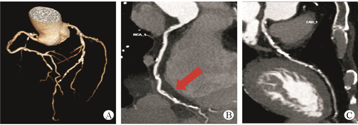
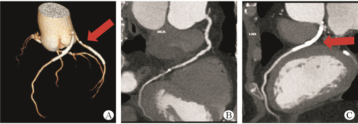
结果(表 2)显示:屏气组与自由呼吸组患者主动脉的SNR差异无统计学意义;屏气组与自由呼吸组RCA近段及中段的CNR差异无统计学意义;屏气组和自由呼吸组LCA、LAD、LCX的CNR差异无统计学意义。2组患者的主动脉、RCA、LM密度差异无统计学意义。2名医师测量一致性良好。图 2、图 3分别显示屏气组和自由呼吸组典型患者CCTA情况。
| 指标 | 屏气组(n=47) | 自由呼吸组(n=47) | 总计(n=94) | t/χ2值 | P值 |
| SNR | 23.64±4.27 | 22.85±3.83 | 23.24±4.05 | 0.87 | 0.39 |
| 图像噪声 | 20.41±2.65 | 20.74±1.68 | 20.58±2.21 | -0.70 | 0.46 |
| CNR | |||||
| 右冠脉近段 | 26.24±5.58 | 25.74±4.60 | 25.99±5.09 | 0.44 | 0.66 |
| 右冠脉中段 | 26.54±6.17 | 25.11±4.41 | 25.82±5.38 | 1.19 | 0.23 |
| 左冠脉 | 26.86±4.31 | 25.84±4.08 | 26.35±4.20 | 1.08 | 0.28 |
| 左前降支主干 | 25.67±5.35 | 24.52±4.35 | 25.10±4.88 | 1.05 | 0.30 |
| 左旋支 | 25.31±5.26 | 23.26±3.99 | 24.28±4.75 | 1.97 | 0.05 |
| CT值/HU | |||||
| 主动脉 | 495.54±94.61 | 464.08±87.76 | 479.81±92.08 | 1.59 | 0.34 |
| 右冠脉近段 | 451.95±116.94 | 427.01±96.88 | 439.48±107.48 | 1.08 | 0.28 |
| 左冠脉 | 470.48±92.83 | 437.52±94.44 | 454.00±94.55 | 0.51 | 0.61 |
| SNR:信噪比;CNR:对比噪声比 | |||||

|
| 图 2 屏气状态下患者CCTA影像 男性,65岁,冠脉粥样硬化性心脏病伴高血压近20年,因胸闷、气短加重再次就诊,行CCTA检查,心率为77次/min. A:容积再现(VR)图像显示冠脉多处钙化;B: 右冠脉管壁见多发软斑块及钙化,曲面重组(CPR)显示血管管腔中度狭窄(箭头处);C:左前降支多发钙化,中段局部管腔中重度狭窄,余管腔显示清晰 |

|
| 图 3 自由呼吸状态下患者CCTA影像 男性,58岁,2017年因冠状动脉狭窄行冠脉支架植入术,2020年11月冠脉支架植入术后复查CCTA,此次检查中心率为72次/min.A:容积再现(VR)图像显示清晰,左前降支、左旋支支架显示清晰;B:右冠中段散在钙化,管腔狭窄小于50%;C:曲面重组(CPR)显示血管管腔通畅,支架边缘光滑锐利(箭头处) |
屏气组包含47支右冠脉(RCA)、46支右后降支(R-PDA)、45支右后外侧支(R-PLB)、47支左冠脉主干(LM)、47支左前降支(LAD)、46支第一对角支(D1)、45支第二对角支(D2)、47支左旋支(LCX)、39支第一钝缘支(OM1)、9支第二钝缘支(OM2)、5支左后外侧支(L-PLB)、13支冠脉分支(RAMUS),各分支评分见表 3。自由呼吸组包含45支RCA、46支R-PDA、45支R-PLB、47支LM、47支LAD、46支D1、45支D2、47支LCX、39支OM1、9支OM2、5支L-PLB、13支RAMUS,各分支评分见表 4。两组患者图像由放射科医生评价,一致性良好(κ = 0.77,P < 0.05)。
| n=47 | |||||||||||||||||||||||||||||
| 部位 | 评分n(%) | 平均分值 | |||||||||||||||||||||||||||
| 5分 | 4分 | 3分 | 2分 | 1分 | |||||||||||||||||||||||||
| 右冠脉 | |||||||||||||||||||||||||||||
| 近段 | 38(80.9) | 7(14.9) | 2(4.2) | 0 | 0 | 4.8±0.5 | |||||||||||||||||||||||
| 中段 | 39(83.0) | 4(8.5) | 4(8.5) | 0 | 0 | 4.7±0.6 | |||||||||||||||||||||||
| 远段 | 39(83.0) | 6(12.8) | 1(2.1) | 1(2.1) | 0 | 4.8±0.6 | |||||||||||||||||||||||
| 右后降支 | 40(87.0) | 2(4.3) | 4(8.7) | 0 | 0 | 4.8±0.6 | |||||||||||||||||||||||
| 右后外侧支 | 41(91.2) | 2(4.4) | 2(4.4) | 0 | 0 | 4.9±0.1 | |||||||||||||||||||||||
| 左冠脉主干 | 46(97.9) | 1(2.1) | 0 | 0 | 0 | 4.9±0.1 | |||||||||||||||||||||||
| 左前降支 | |||||||||||||||||||||||||||||
| 近段 | 46(97.9) | 1(2.1) | 0 | 0 | 0 | 4.9±0.1 | |||||||||||||||||||||||
| 中段 | 39(83.0) | 7(14.9) | 1(2.1) | 0 | 0 | 4.8±0.4 | |||||||||||||||||||||||
| 远段 | 35(74.6) | 6(12.8) | 4(8.3) | 2(4.3) | 0 | 4.6±0.8 | |||||||||||||||||||||||
| 第一对角支 | 3(6.5) | 32(69.6) | 9(19.6) | 2(4.3) | 0 | 3.8±0.6 | |||||||||||||||||||||||
| 第二对角支 | 6(13.3) | 8(17.8) | 18(40.0) | 13(28.9) | 0 | 3.2±1.0 | |||||||||||||||||||||||
| 左旋支 | |||||||||||||||||||||||||||||
| 近段 | 45(95.7) | 2(4.3) | 0 | 0 | 0 | 4.9±0.2 | |||||||||||||||||||||||
| 中段 | 40(85.1) | 5(10.6) | 2(4.3) | 0 | 0 | 4.9±0.4 | |||||||||||||||||||||||
| 远段 | 22(46.8) | 13(27.7) | 10(21.3) | 2(4.3) | 0 | 4.0±0.9 | |||||||||||||||||||||||
| 第一钝缘支 | 11(28.2) | 14(35.9) | 12(30.8) | 2(5.1) | 0 | 4.0±0.9 | |||||||||||||||||||||||
| 第二钝缘支 | 2(22.2) | 5(55.6) | 2(22.2) | 0 | 0 | 3.9±0.6 | |||||||||||||||||||||||
| 左后外侧支 | 2(40.0) | 3(50.0) | 0 | 0 | 0 | 4.3±0.7 | |||||||||||||||||||||||
| 冠脉分支 | 0 | 11(84.6) | 2(15.6) | 0 | 0 | 3.9±0.7 | |||||||||||||||||||||||
| n=47 | |||||||||||||||||||||||||||||
| 部位 | 评分n(%) | 平均分值 | |||||||||||||||||||||||||||
| 5分 | 4分 | 3分 | 2分 | 1分 | |||||||||||||||||||||||||
| 右冠脉 | |||||||||||||||||||||||||||||
| 近段 | 36(80.0) | 9(20.0) | 0 | 0 | 0 | 4.8±0.4 | |||||||||||||||||||||||
| 中段 | 41(91.1) | 4(8.9) | 0 | 0 | 0 | 4.9±0.3 | |||||||||||||||||||||||
| 远段 | 42(93.3) | 3(0.67) | 0 | 0 | 0 | 4.9±0.2 | |||||||||||||||||||||||
| 右后降支 | 24(58.5) | 12(29.3) | 4(9.8) | 1(2.4) | 0 | 4.4±0.8 | |||||||||||||||||||||||
| 右后外侧支 | 28(66.7) | 12(28.6) | 2(4.8) | 0 | 0 | 4.6±0.6 | |||||||||||||||||||||||
| 左冠脉主干 | 43(95.6) | 2(4.4) | 0 | 0 | 0 | 4.9±0.2 | |||||||||||||||||||||||
| 左前降支 | |||||||||||||||||||||||||||||
| 近段 | 39(86.7) | 5(11.1) | 1(2.2) | 0 | 0 | 4.8±0.4 | |||||||||||||||||||||||
| 中段 | 41(91.1) | 4(8.9) | 0 | 0 | 0 | 4.9±0.3 | |||||||||||||||||||||||
| 远段 | 37(82.2) | 7(15.6) | 1(2.2) | 2(4.3) | 0 | 4.8±0.5 | |||||||||||||||||||||||
| 第一对角支 | 4(8.9) | 27(60.0) | 13(28.9) | 1(2.2) | 0 | 3.8±0.6 | |||||||||||||||||||||||
| 第二对角支 | 5(12.5) | 17(42.5) | 17(42.5) | 5(12.5) | 0 | 3.5±0.9 | |||||||||||||||||||||||
| 左旋支 | |||||||||||||||||||||||||||||
| 近段 | 43(95.6) | 0 | 0 | 0 | 0 | 4.9±0.2 | |||||||||||||||||||||||
| 中段 | 42(93.4) | 1(2.2) | 1(2.2) | 0 | 0 | 4.9±0.4 | |||||||||||||||||||||||
| 远段 | 20(44.4) | 15(33.4) | 15(33.4) | 2(4.4) | 0 | 4.0±0.9 | |||||||||||||||||||||||
| 第一钝缘支 | 14(36.0) | 10(25.6) | 10(25.6) | 2(5.1) | 0 | 4.0±0.9 | |||||||||||||||||||||||
| 第二钝缘支 | 1(14.3) | 2(28.6) | 2(28.6) | 0 | 0 | 3.9±0.6 | |||||||||||||||||||||||
| 左后外侧支 | 1(14.3) | 1(14.2) | 1(14.2) | 0 | 0 | 4.3±0.7 | |||||||||||||||||||||||
| 冠脉分支 | 4(18.2) | 4(18.2) | 4(18.2) | 1(4.5) | 0 | 3.9±0.7 | |||||||||||||||||||||||
屏气组总评分段数为718段,自由呼吸组总评分段数为693段。评分在3分以上符合诊断要求。屏气组3.1%的图像不符合诊断要求,自由呼吸组1.9%的图像不符合诊断要求,两组图像质量差异无统计学意义(P=0.768,表 5)。
| n(%) | |||||||||||||||||||||||||||||
| 评分 | 屏气组(n=718) | 自由呼吸组(n=693) | 总计 | ||||||||||||||||||||||||||
| 5分 | 494(68.8) | 467(67.4) | 961(68.1) | ||||||||||||||||||||||||||
| 4分 | 129(18.0) | 141(20.3) | 270(19.1) | ||||||||||||||||||||||||||
| 3分 | 73(10.2) | 72(10.4) | 145(10.3) | ||||||||||||||||||||||||||
| 2分 | 22(3.1) | 13(1.9) | 35(2.5) | ||||||||||||||||||||||||||
| 1分 | 0 | 0 | 0 | ||||||||||||||||||||||||||
本研究基于320排探测器CT在患者自由呼吸或屏气状态下对其进行CCTA,比较两组患者的辐射剂量和图像质量,探究心率 < 75次/min患者行CCTA自由呼吸的可行性。本研究采用首台国产320排CT(联影),探测器宽度为16 cm,球管旋转时间也达到0.25 s,并对此机器成像性能进行探讨。结果表明,自由呼吸和屏气患者的SNR和CNR差异无统计学意义,说明心率 < 75次/min患者行CCTA时不受自由呼吸影响,与近年来多项相关研究[12-17]结果一致。
以往研究[18]发现,正常呼吸时(12~20次/min),膈肌运动引起的冠脉运动速度为6.4~29.3 mm/s,心跳引起的冠脉速度为22.4~108.6 mm/s,提示呼吸引起的伪影可忽略不计。宽探测器CT的发展有助于提高时间分辨率,从而可抑制冠脉的运动伪影,包括由心脏运动和呼吸运动引起的运动伪影[19]。与屏气相比,自由呼吸CCTA减少了屏气指令引起的心率变异,进而提高了成功率[20]。uCT960+是目前转速最快的320排CT,将超高系统转速与时空探测器结合,实现了时间分辨率、空间分辨率与对比度分辨率的三重提升。ePhase智能寻心算法,基于各心动周期智能重建和选择最佳时相,能提升复杂患者的检查成功率。本研究中宽探测器CT共观察了1 411个冠脉节段,其中屏气组718个中,696个(96.9%)可用于诊断;自由呼吸组693个冠脉节段中,680个(98.1%)可用于诊断。两组图像质量差异无统计学意义,与以往研究结果[21-24]相近。由于宽体探测器CT宽度达16 cm,时间分辨率为0.25 s,可在1个心动周期内旋转1周而获得全心脏数据,减少冠脉运动伪影。不同扫描参数之间相互切换时间也得以缩短,可在CCTA成像时间点瞬时切换成相对较高的参数。此外,宽探测器CT也明显缩短了曝光时间、成像时间,因此对比剂用量和ED较少(P < 0.01)。
综上所述,对于心率 < 75次/min患者,自由呼吸CCTA是一种可以接受的方案。本研究的不足之处在于未研究同一患者自由呼吸和屏气时的CCTA图像,未将冠脉造影结果作为金标准等,后续将继续深入研究。
利益冲突:所有作者声明不存在利益冲突
| [1] |
KANG E J, LEE J, LEE K N, et al. An initial randomised study assessing free-breathing CCTA using 320-detector CT[J]. Eur Radiol, 2013, 23(5): 1199-1209.
[DOI]
|
| [2] |
KINO A, ZUCKER E J, HONKANEN A, et al. Ultrafast pediatric chest computed tomography: comparison of free-breathing vs. breath-hold imaging with and without anesthesia in young children[J]. Pediatr Radiol, 2019, 49(3): 301-307.
[DOI]
|
| [3] |
SHUAI T, DENG L, PAN Y, et al. Free-breathing coronary CT angiography using 16-cm wide-detector for challenging patients: comparison with invasive coronary angiography[J]. Clin Radiol, 2018, 73(11): 986-987.
[PubMed]
|
| [4] |
LIU Z, SUN Y, ZHANG Z, et al. Feasibility of free-breathing CCTA using 256-MDCT[J]. Medicine, 2016, 95(27): 4096-4098.
[DOI]
|
| [5] |
GHEKIERE O, SALGADO R, BULS N, et al. Image quality in coronary CT angiography: challenges and technical solutions[J]. Br J Radiol, 2017, 90(10): 2567-2569.
[PubMed]
|
| [6] |
COLLET C, ONUMA Y, ANDREINI D, et al. Coronary computed tomography angiography for heart team decision-making in multivessel coronary artery disease[J]. Eur Heart J, 2018, 39(15): 352-356.
[PubMed]
|
| [7] |
CAO L, LIU X, LI J Y, et al. Improving the degree and uniformity of enhancement in coronary CT angiography with a new bolus tracking method enabled by free breathing[J]. Acad Radiol, 2019, 26(12): 1591-1596.
[DOI]
|
| [8] |
ZREIK M, HAMERSVELT R V, WOLTERINK J M, et al. A recurrent CNN for automatic detection and classification of coronary artery plaque and stenosis in coronary CT angiography[J]. IEEE Trans Med Imaging, 2018, 17(21): 224-226.
[URI]
|
| [9] |
DI CESARE E, GENNARELLI A, DI SIBIO A, et al. Image quality and radiation dose of single heartbeat 640-slice coronary CT angiography: a comparison between patients with chronic Atrial Fibrillation and subjects in normal sinus rhythm by propensity analysis[J]. Eur J Radiol, 2015, 84(4): 631-636.
[DOI]
|
| [10] |
GOO H W. Quantitative evaluation of coronary artery visibility on CT angiography in Kawasaki disease: young vs. old children[J]. Int J Cardiovasc Imaging, 2020, 16(7): 331-335.
[DOI]
|
| [11] |
MUSHTAQ S, CONTE E, PONTONE G, et al. Interpretability of coronary CT angiography performed with a novel whole-heart coverage high-definition CT scanner in 300 consecutive patients with coronary artery bypass grafts[J]. J Cardiovasc Comput Tomogr, 2020, 14(2): 137-143.
[DOI]
|
| [12] |
JOHNSON K M, JOHNSON H E, YANG Z, et al. Scoring of coronary artery disease characteristics on coronary ct angiograms by using machine learning[J]. Radiology, 2019, 292(2): 182-185.
[URI]
|
| [13] |
ANDREINI D, PONTONE G, MUSHTAQ S, et al. Atrial fibrillation: diagnostic accuracy of coronary CT angiography performed with a whole-heart 230-μm spatial resolution CT scanner[J]. Radiology, 2017, 284(3): 676-684.
[DOI]
|
| [14] |
LIANG J, WANG H, XU L, et al. Diagnostic performance of 256-row detector coronary CT angiography in patients with high heart rates within a single cardiac cycle a preliminary study[J]. Clin Radiol, 2017, 72(8): 694-695.
[URI]
|
| [15] |
ZHU Y, LI Z, MA J, HONG Y, et al. Imaging the infant chest without sedation: feasibility of using single axial rotation with 16-cm wide-detector CT[J]. Radiology, 2018, 286(1): 279-285.
[DOI]
|
| [16] |
SCHOLTZ J E, ADDISON D, BITTNER D O, et al. Diagnostic Performance of coronary CTA in intermediate-to-high-risk patients for suspected acute coronary syndrome[J]. Jacc Cardiovascular Imaging, 2018, 51(12): 193-195.
[URI]
|
| [17] |
LI Z N, YIN W H, LU B, et al. Improvement of image quality and diagnostic performance by an innovative motion-correction algorithm for prospectively ECG triggered coronary CT angiography[J]. PLoS One, 2015, 10(11): 142-145.
[URI]
|
| [18] |
SONG I, KANG J H, KIM M Y, et al. Diagnostic accuracy of electrocardiogram-gated thoracic computed tomography angiography without heart rate control for detection of significant coronary artery stenosis in patients with acute ischemic stroke: a comparative study[J]. Korean J Radiol, 2018, 19(5): 905-915.
[DOI]
|
| [19] |
LIU Z, ZHANG Z, HONG N, et al. Diagnostic performance of free-breathing coronary computed tomography angiography without heart rate control using 16-cm z-coverage CT with motion-correction algorithm[J]. J Cardiovasc Comput Tomogr, 2019, 13(2): 113-117.
[DOI]
|
| [20] |
GHANEM A M, HAMIMI A H, GHARID A M, et al. Automatic assessment of 3D coronary artery distensibility from time-resolved coronary CT angiography[C]. Ann Int Conf IEEE Eng Med Biol Soc, 2019, 2019: 836-840.
|
| [21] |
LU M T, FERENCIK M, ROBERTS R S, et al. Noninvasive FFR derived from coronary CT angiography: management and outcomes in the PROMISE Trial[J]. JACC Cardiovasc Imaging, 2017, 11(8): 369-372.
[URI]
|
| [22] |
CHEN Y, WEI D, LI D, et al. The value of 16-cm wide-detector computed tomography in coronary computed tomography angiography for patients with high heart rate variability[J]. J Comput Assist Tomogr, 2018, 42(6): 906-911.
[DOI]
|
| [23] |
LI Y, CAO X, XING Z, et al. MO-AB-BRA-10:super high temporal resolution cardiac CT imaging using SMART-RECON[J]. Med Phys, 2016, 43(6): 3692-3693.
[URI]
|
| [24] |
FUNABASHI N, NAMIHIRA Y, IRIE R, et al. Multisector-reconstruction in 1st generation 320-slice CT at high pulsation-rates achieved accurate-evaluation of coronary-lumen patency after insertion of a XIENCE stent. XIENCE Phantom Study Part 4[J]. Int J Cardiol, 2016, 20(2): 509-510.
[URI]
|
 2021, Vol. 28


